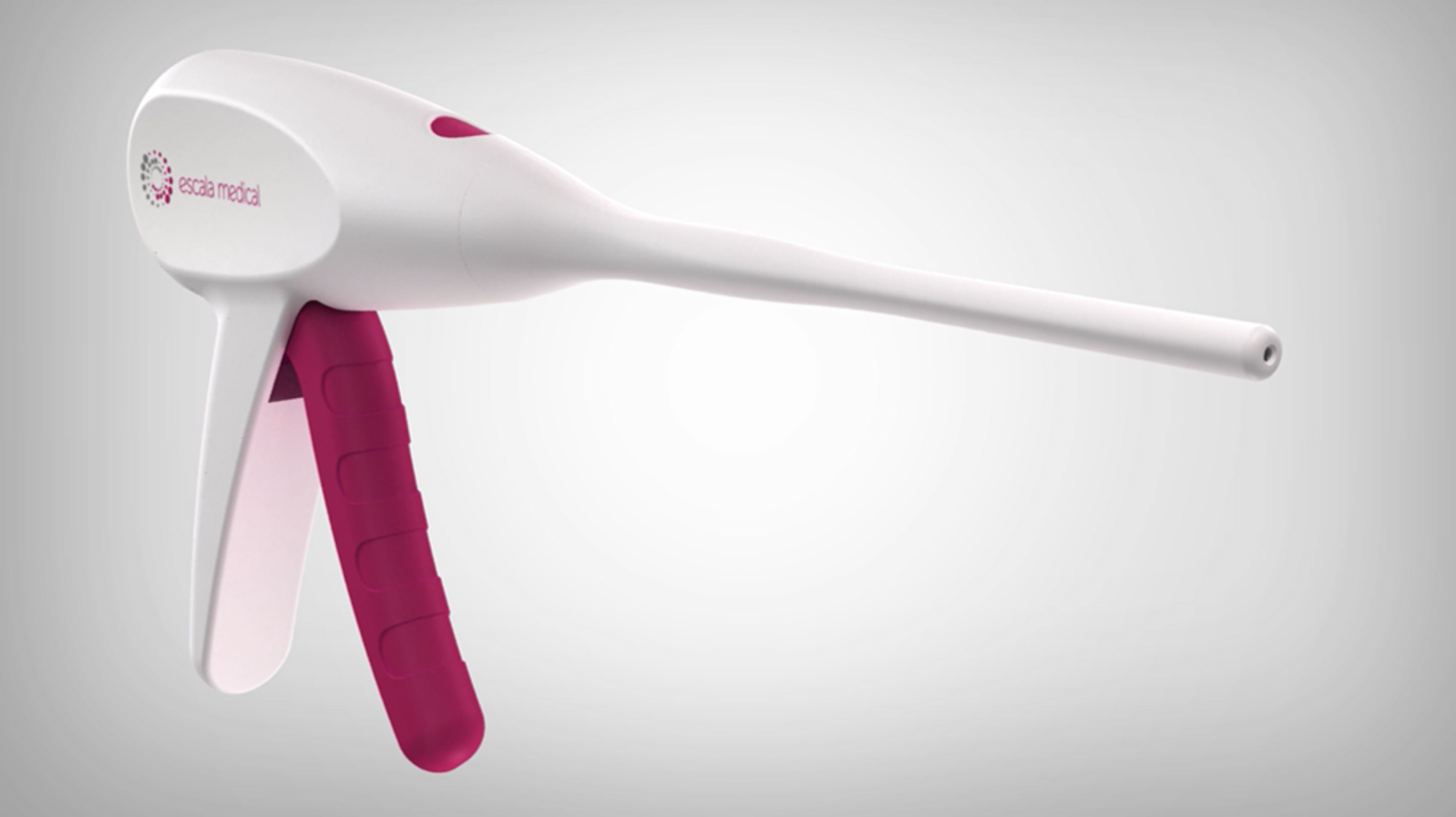
Unfortunately, this ridiculous looking device will soon be inserted deeply into the pelvic cavity of countless women to staple the vagina to the sacrospinous ligament, a new twist on an old operation fraught with risk and failure.
The device is being ushered to market using the Food and Drug Administration’s 510K process, whereby safety and efficacy studies are automatically waved based on established and equivalent procedures, in this case the sacrospinous ligament fixation (SSLF).
Developed by vaginal surgeons Nichols and Randall in 1971, until recently SSLF has been an operation reserved exclusively for post-hysterectomy women. During SSLF a midline incision is made in the posterior vaginal wall. Blunt digital dissection is extended deeply behind the rectum and into the coccygeus muscle of the pelvic wall. Connective tissue overlying the coccygeus muscle is removed to completely expose the sacrospinous ligament. Sutures are then placed in both the ligament and top of the vagina. Since the vaginal stump is not long enough to reach both ligaments, unilateral fixation is the standard practice.
 SSLF has always been associated with anatomic distortion of the vagina and rectum, chronic pain, and severe and intractable postoperative cystocele. In normal anatomy the uterus pulls the vagina toward the front of the body. As the bladder and uterus are connected at the level of the cervix, the bladder is also pulled forward into its normal position against the lower abdominal wall.
The ill-conceived SSLF pulls the vagina in the opposite direction by tethering it to the back of the body, thus pulling the bladder backward against the front vaginal wall. Sexual disability and bowel dysfunction are also well-known risk factors. The pudendal nerve, embedded between the sacrospinous and sacrotuberous ligaments, can easily be damaged by this surgery, leading to chronic pain and anal sphincter dysfunction. Not a single study in all of gynecologic or orthopedic literature describes the musculoskeletal risks of having the muscular vagina tethered to one side of the body.
Bilateral SSLF without hysterectomy has recently been described, using strips of polypropylene mesh to bridge the gap between cervix and both sacrospinous ligaments. Erosion and exposure of the mesh has been reported in up to 37.5% of subjects during short-term follow up studies*. Often sold as minimally invasive, it is nothing less than criminal malpractice to surgically tether the normally anteverted uterus to the back of the body.
Although Escala Medical states it will “Soon offer an alternative first-line solution for milder cases of prolapse and can help achieve a better outcome in more severe conditions – especially for women who aren’t good surgical candidates or don’t want surgery”, the anatomic realities of SSLF suggest this device will be used post-hysterectomy. Therefore, hysterectomy will likely be offered as part of the operation.
Many pelvic surgeons have abandoned SSLF in favor of abdominal sacrocolpopexy, which tethers the vaginal stump to the anterior ligament of the second sacral vertebra by way of a mesh bridge. A difficult operation also associated with risk and failure, at least sacrocolpopexy results in a more natural vaginal axis and therefore fewer postoperative complications.
Escala tells us, “The potential market adds up to $600 million in the United States alone, where close to 400,000 prolapse repair procedures are performed annually at a direct cost of $1.4 billion.”
It is a very dark side of human nature that pursuit of money often blinds otherwise caring and ethical human beings into the swamp of surgical opportunity and allows them to ignore the lifelong damage done to the patients, especially women.
___________________
* Senturk M Guraslan H Cakmak Y Ekin M Bilateral sacrospinous fixation without hysterectomy: 18-month follow up. Journal of the Turkish-German Gynecological Association. 16(2): 102-106 2015


